 Electromyography Electromyography
The term Electromyography (EMG) encompasses nerve conduction studies and the needle exam. Both of these are discussed below:
The nerve conduction study (NCS), is often performed at the same time with the same equipment. In this test, stimulating and recording electrodes are used, and small electrical shocks are applied to measure the ability of the nerve to conduct electrical signals. This test may cause mild tingling and discomfort similar to a mild shock from static electricity. Nerve conduction velocity and evoked potential testing are especially helpful when pain or sensory complaints are more prominent than weakness. (For more information on NCS, click here.)
The needle exam portion is an electrical recording of muscle activity that aids in the diagnosis of neuromuscular disease.
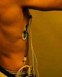
During an EMG test, a fine needle is inserted into the muscle to be tested. This may cause some discomfort, similar to that of an injection. Recordings are made while the muscle is at rest, and then during the contraction. The person performing the test may move the limb being tested, and direct the patient to move it with various levels of force. A typical session lasts from 20-30 minutes.
Though the needle is placed in a muscle, the recording tells Dr. Nelson as much about the nerve as it does the muscle. EMG is performed most often to help diagnose different diseases causing weakness and identify abnormalities of nerves or spinal nerve roots that may be associated with pain or numbness. EMG results can help determine whether symptoms are due to a muscle disease or a neurological disorder, and, when combined with clinical findings, usually allow a confident diagnosis. EMG can help diagnose many muscle and nerve disorders including muscular dystrophy, myopathies, peripheral neuropathies, nerve lesions and spinal muscular atrophy. (For more information regarding the needle portion of EMG, click here.)
If an EMG has been ordered for you, take all of your normal medications as directed and do not use skin lotion on the day of your study.
Electroencephalography (EEG)
Electroencephalography (EEG) is the diagnostic field devoted to testing the central nervous system. It involves the recording and evaluation of spontaneous evoked electrical activity from the brain through surface disk electrodes attached to your scalp with a conductive, adhesive cream. During the test, you will lie in a prone position. The test is totally non-invasive. Every effort is made to make you comfortable and relaxed in a quiet environment. During the recording stage of your test, you will be asked to complete various tasks, such as opening and closing your eyes, or hyperventilating for about four minutes.
Also known as a brain wave test, it is a key tool in the diagnosis and management of epilepsy and other seizure disorders. It is also used to assist in the diagnosis of brain damage and diseases such as strokes, tumors, encephalitis, mental retardation, and sleep disorders. The results of the test can also distinguish some psychiatric conditions from degenerative mental disorders such as Alzheimer's and Parkinson's diseases.
If you are scheduled for an EEG, please take your routine medicines on the day of the test and avoid caffeine.
|